



 Pilhan Kim, CEO
However, preclinical IVM studies face challenges in achieving high-resolution imaging for small molecules, maintaining imaging setup stability without causing harm from phototoxicity, and managing large and complex imaging data from two-photon and confocal systems. The successful implementation of these studies demands expertise in specialized data analysis techniques.
Our All-in-One Intravital Confocal/Two-Photon microscopy system is extensively optimized and carefully engineered to provide superb performance in the intravital imaging of live animal models in vivo
Given the ethical considerations surrounding animal welfare, strict adherence to ethical guidelines is paramount to mitigate any harm or distress to live animals involved in IVM studies.
“Our All-in-One Intravital Confocal/Two-Photon microscopy system is extensively optimized and carefully engineered to provide superb performance in the intravital imaging of live animal models in vivo. It facilitates the assessment of drug delivery efficiency for antibodies, exosomes, organic compounds, or nanoparticle-based drug delivery in any target organ or tissue, making it a versatile tool for preclinical research and development,” says Pilhan Kim, CEO of IVIM Technology.
In the capable hands of its preclinical research and development team, IVIM’s intravital microscopes offer a remarkable ability to image virtually any organ or tissue within a living mouse. Capabilities extend to directly visualizing the pharmacokinetics and mechanism/ pathophysiology of drugs and biopharmaceutical compounds. This includes in vivo drug delivery and efficacy monitoring across various models, such as primary/metastatic cancer, normal or eye disease in the retina and cornea, and normal and brain disease mouse models.
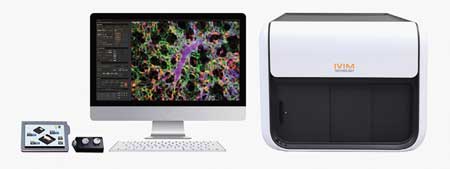
IVIM’s systems enable the study of cell movement in living tissues, providing valuable information about individual cell dynamics in different organs. The technology operates at both the cellular and subcellular levels, detecting complex relationships between biological elements such as muscle, adipose tissue, proteins, and exosomes in their microenvironments. IVIM’s solutions deliver superior speed, resolution, and imaging precision, utilizing user-friendly software, animal motion compensation, AI-based noise cancellation and processing, optimized imaging setups, licensed imaging windows, and proprietary methodologies supported by expertise for an exceptional imaging experience.
IVIM’s intravital in vivo imaging technology revolutionizes the field by enabling extended, long-term observation of disease and injury development within the target organ. An outstanding example is its ability to perform continuous in vivo imaging of cancer xenograft models, extending from one week to several months. IVIM’s innovative approach, using optimized imaging techniques and dorsal skinfold chambers, allows researchers to observe phenomena previously impossible with traditional methods. This advancement holds the potential to significantly enhance the understanding of disease progression and therapeutic responses in the study of human diseases.
Illustrating IVIM’s impact, it provided CRO services to a leading global exosome company. The research involved in vivo imaging to study the involvement of exosomes in systemic inflammation within septic mouse models. Additionally, it focused on examining the biodistribution of these exosomes in various organs like the brain, spleen, and liver or specific parts in the organ like the blood-brain barrier in the brain. This approach allowed for a clearer understanding of the mode of action necessary for new drug development, surpassing in vitro studies with more reliable in vivo results. Consequently, the client’s drug development timeline was significantly reduced.
IVIM remains at the forefront of innovation through continuous research and development efforts. It proudly offers both online and onsite demonstrations, customer training, as well as regular seminars to keep its clients informed and engaged.
